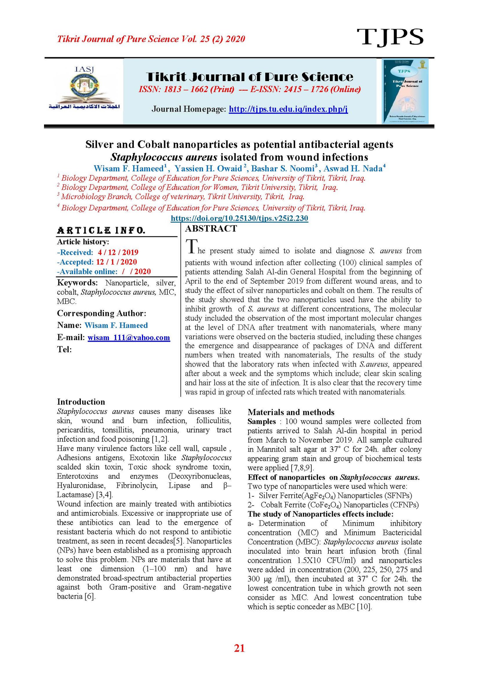
Silver and Cobalt nanoparticles as potential antibacterial agents Staphylococcus aureus isolated from wound infections
Main Article Content
Abstract
The present study aimed to isolate and diagnose S. aureus from patients with wound infection after collecting (100) clinical samples of patients attending Salah Al-din General Hospital from the beginning of April to the end of September 2019 from different wound areas, and to study the effect of silver nanoparticles and cobalt on them. The results of the study showed that the two nanoparticles used have the ability to inhibit growth of S. aureus at different concentrations, The molecular study included the observation of the most important molecular changes at the level of DNA after treatment with nanomaterials, where many variations were observed on the bacteria studied, including these changes the emergence and disappearance of packages of DNA and different numbers when treated with nanomaterials, The results of the study showed that the laboratory rats when infected with S.aureus, appeared after about a week and the symptoms which include; clear skin scaling and hair loss at the site of infection. It is also clear that the recovery time was rapid in group of infected rats which treated with nanomaterials
Article Details

This work is licensed under a Creative Commons Attribution 4.0 International License.
Tikrit Journal of Pure Science is licensed under the Creative Commons Attribution 4.0 International License, which allows users to copy, create extracts, abstracts, and new works from the article, alter and revise the article, and make commercial use of the article (including reuse and/or resale of the article by commercial entities), provided the user gives appropriate credit (with a link to the formal publication through the relevant DOI), provides a link to the license, indicates if changes were made, and the licensor is not represented as endorsing the use made of the work. The authors hold the copyright for their published work on the Tikrit J. Pure Sci. website, while Tikrit J. Pure Sci. is responsible for appreciate citation of their work, which is released under CC-BY-4.0, enabling the unrestricted use, distribution, and reproduction of an article in any medium, provided that the original work is properly cited.
References
[1] Khudaier, B.Y.; Abbas, B.A. and Khudaier, A. M. (2013). Detection of Methicillin Resistant S.aureus Isolated from Human and Animals in Basrah Province /Iraq. Mirror of Research in Veterinary Sciences and animals. MRSVA 2 (3):12-21.
[2] Abdul Aziz, J. M. and Kalavathi, F. (2017). Prevalence of methicillin resistant S.aureus isolates and their antibiotic susceptibility in tertiary care hospitals, India. Edorium Journal Microbial, 3:18–23. [3]- Hallabjaiy, R.; Darogha, S. N. and Hamad, P. A. (2014). Vancomycin Resistance Among Methicillin-Resistant S.aureus Isolated from Clinical Samples in Erbil City-Iraq. Medical Journal of Islamic World Academy of Sciences,109 (1646):1-7.
[4] Garba, S.; Igwe, J.C.; Onaolapo, J.A. and Olayinka, B.O. (2018). Vancomycin Resistant S.aureus from Clinical Isolates in Zaria Metropolis, Kaduna State. Clinical Infect Dis 2: 105.
[5] Fricke, E.C.; Haak, D.C.; Levey, D.J. and Tewksbury, J.J. (2016). Gut passage and secondary metabolites alter the source of post-dispersal predation for bird-dispersed chili seeds. Ecologies, 181(3): 905-910.
[6] El-Bayomi, R. M.; Ahmed, H. A.; Awadallah, M. A., Mohsen, R. A., Abd El-Ghafar, A. E.; and Abdelrahman, M. A. (2016). Occurrence, Virulence Factors, Antimicrobial Resistance, and Genotyping of Staphylococcus aureus Strains Isolated from Chicken Products and Humans. Vector Borne Zoonotic Dis, 16(3): 157-164.
[7] Axtner J, Crampton-Platt A, Hörig LA, Mohamed A, Xu CCY, Yu DW. (2019).Wilting A.Gigascience. An efficient and robust laboratory workflow and tetrapod database for larger scale environmental DNA studies.. Apr 8(4): 1.
[8] Chan, C. L.; Gillbert, A.; Basuino, L. Hamitton, K. and Chatterjee, S. S. (2016). PBP4 mediates high-level resistance to new generation cephalosporins in Staphylococcus aureus. Journal. Antimicrob. Agents Chemother.ACC.00358-16: 1-24.
[9] Singh G. K.; Bopanna B. D.; and Rindhe G. (2014). Molecular characterization of Staphylococcus aureus - human pathogen from clinical samples by RAPD markers, Int. Journal. of Curr .Microbiol. App. Sci., 3(2): 349-354.
[10] Akanbi, O.E.; Njom, H.A.; Fri, J., Otigbu, A.O. and Clarke, A.M.(2017). Antimicrobial Susceptibility of S.aureus Isolated from Recreational Waters and Beach Sand in Eastern Cape Province of South Africa. Int. Journal. Environ. Res. Public Health, 14: 1001. [11] Onasanya, A., Mignouna ,H.D. &Thottappilly, G.(2003). Genetic fingerprinting and phylogenetic diversity of Staphylococcus aureus isolates from Nigeria. African journal of biotechnology, 2(8): 246-250.
[12] Afrough P. ; Pourmand M. R.; Sarajian A. A.; Saki M. and Saremy S.(2013). Molecular Investigation of Staphylococcus aureus, coa and spa Genes in Ahvas Hospitals, Staff Nose Compared With Patients Clinical Samples, Jundishapur Journal. Microbiol, 6(4): 1-7.
[13] Sharma,p.; Sharma, A.; Sharma,M.; Bhalla,N.; Estrela,P.; Jain,A.; Thakur, and Thakur, A.(2017). Nanomaterial Fungicides: In Vitro and In Vivo Antimitotic Activity of Cobalt and Nickel Nano ferrites on Phyto pathogenic Fungi. WILEY-VCH Verlag Gmb H and Co. KGaA, Weinhiem
[14] Amarakoon A. (2016). Detection of C677T and A1298C mutations within the MTHFR gene by PCR and RFLP assays and assessment of risk factor of Hyperhomocysteinemia. WSN 53(3) 253-274 .Marine Biological Resources Division, National Aquatic Resources and Development Agency (NARA), No15, Crow Island, Colombo 15, Sri Lanka.
[15] Edward,A.; Qin Dai ,S.; Kim M.; David M.H.; and Warren C.W.(2014). Nanoparticle Exposure in Animals can be Visualized in the Skin and Analyzed via Skin Biopsy. Canadian Institute of Health Research.
[16] Sorkh, M. A.; Shokoohizadeh, L.; Rashidi, N. and Tajbakhsh, E. (2017). Molecular Analysis of Pseudomonas aeruginosa Strains Isolated from Burn Patients by Repetitive Estrogenic Palindromic-PCR (rep-PCR). Iranian Red Crescent Medical Journal, 19(4).
