Estimation of Furosemide Spectrophotometrically in Pharmaceutical preparations by Oxidative Coupling Reaction
Article Sidebar
Main Article Content
Abstract
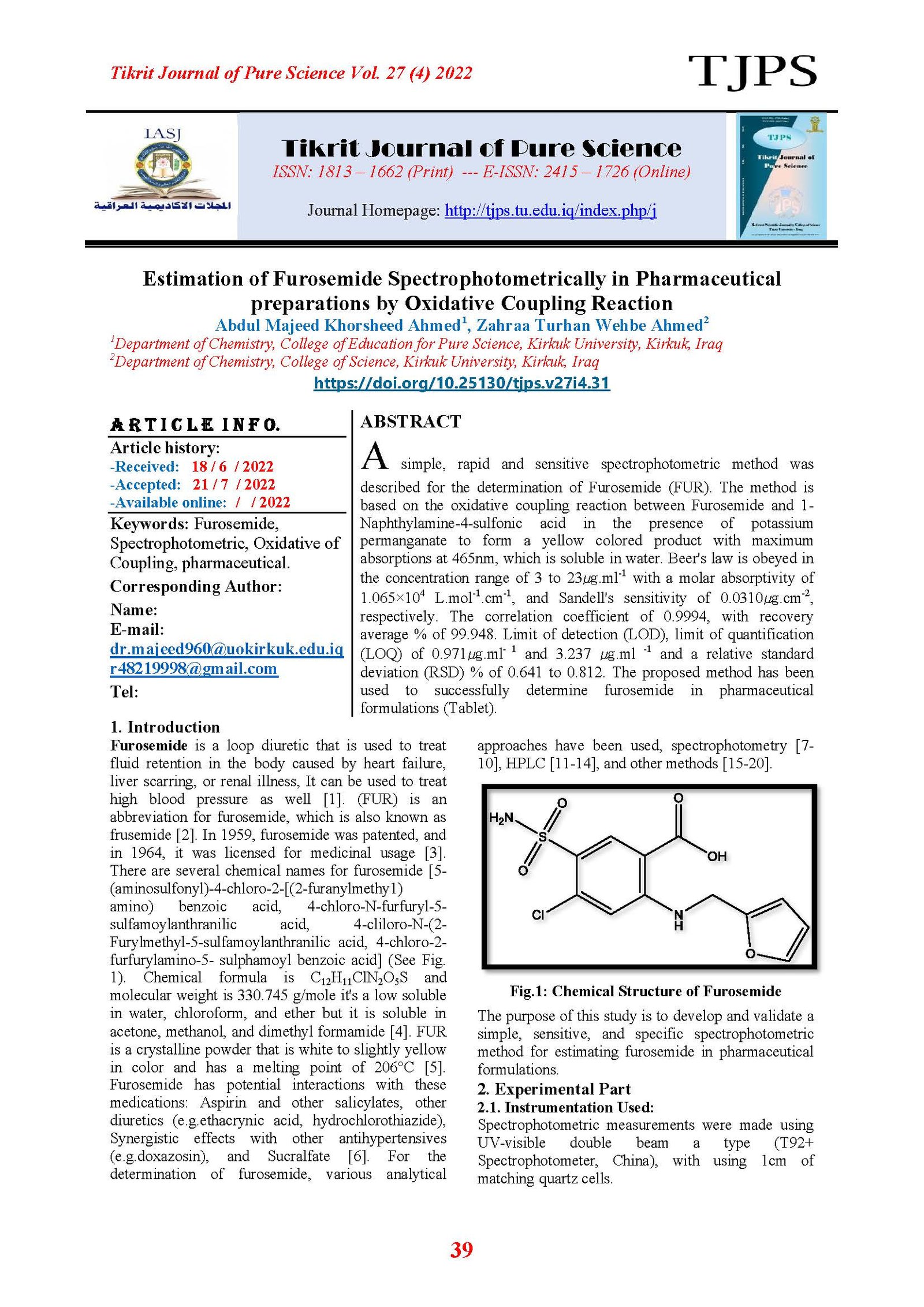
A simple, rapid and sensitive spectrophotometric method was described for the determination of Furosemide (FUR). The method is based on the oxidative coupling reaction between Furosemide and 1-Naphthylamine-4-sulfonic acid in the presence of potassium permanganate to form a yellow colored product with maximum absorptions at 465nm, which is soluble in water. Beer's law is obeyed in the concentration range of 3 to 23㎍.ml-1 with a molar absorptivity of 1.065×10⁴ L.mol-1.cm-1, and Sandell's sensitivity of 0.0310㎍.cm-2, respectively. The correlation coefficient of 0.9994, with recovery average % of 99.948. Limit of detection (LOD), limit of quantification (LOQ) of 0.971㎍.ml- 1 and 3.237 ㎍.ml -1 and a relative standard deviation (RSD) % of 0.641 to 0.812. The proposed method has been used to successfully determine furosemide in pharmaceutical formulations (Tablet).
Article Details

This work is licensed under a Creative Commons Attribution 4.0 International License.
Tikrit Journal of Pure Science is licensed under the Creative Commons Attribution 4.0 International License, which allows users to copy, create extracts, abstracts, and new works from the article, alter and revise the article, and make commercial use of the article (including reuse and/or resale of the article by commercial entities), provided the user gives appropriate credit (with a link to the formal publication through the relevant DOI), provides a link to the license, indicates if changes were made, and the licensor is not represented as endorsing the use made of the work. The authors hold the copyright for their published work on the Tikrit J. Pure Sci. website, while Tikrit J. Pure Sci. is responsible for appreciate citation of their work, which is released under CC-BY-4.0, enabling the unrestricted use, distribution, and reproduction of an article in any medium, provided that the original work is properly cited.
References
[1] M. E. Bosch, A. R. Sánchez, F. S. Rojas and C. B. Ojeda, Journal of pharmaceutical and biomedical analysis 48 (3), 519-532, 2008.
[2] B. B. Melane, Thermal and Photostability Studies of Furosemide and Its Cyclodextrin Mixtures, 2002.
[3] F. J. Ganellin CR. Analogue-based Drug Discovery. John Wiley & Sons. p. 458, 2006.ISBN 9783527607495.
[4] A. M. Al-Obaid, F. J. Al-Shammary, K. A. M. Al-Rashood and M. S. Mian, "Analytical profiles of drug substances 18, 153-193 (1990).
[5] G. J. Kher, Saurashtra University, 2011.
[6] R.N Brogden, Heel RC, TM Speight and G.S Avery "Sucralfate. A review of its pharmacodynamic properties and therapeutic use in peptic ulcer disease". Drugs. 27 (3): 194–209, March 1984. Doi: 10.2165/00003495-198427030-00002.
[7] S. A. Darweesh, “Simultaneous Determination of Sulfanilamide and Furosemide by Using Derivative Spectrophotometry,” vol. 29, no. 2, pp. 240–253, 2016.
[8] M. E. Hassouna, “Spectrophotometric Determination of Furosemide Drug in Different Formulations using Schiff’s Bases,” Foresic Res. Criminol. Int. J., vol. 1, no. 6, 2016.
[9] A. F. Qader, N. A. Fakhre, “Spectrofluorometric determination of furosemide in some pharmaceutical product using acriflavine as a reagent”, AIP Conf. Proc., vol.1888, no. September, 2017.
[10] F.K.Omar, H. S. Mahmod. “An indarect spectrophotometric determination of furosemide in phamaceutical preparation" In World Journal of Pharmaceutical Research” ,vol.7, no.16, pp.1655-1661, 2018.
[11] M. S. Kaynak, E. Akgeyik, and S. Sahin, “Development of HPLC Methods for Individual Determination of 20 Active Pharmaceutical Ingredients for Ussing-Chamber Studies”, Current pharmaceutical Analysis, vol. 13, pp. 145–153, 2017.
[12] S. Sharma, M. phale. “Stress degradation studies of furosemide and developmant and validation of siam RP-HPLC method for ITS quantification”, In World Journal of Pharmaceutical Research, vol. 6, no. 5, pp. 906–920, 2017.
[13] J. S. Shaikh, N. N. Rao. “Simultaneous estimation and forced degradation studies of amiloride hydrochloride and furosemide in a pharmaceutical dosage form using reverse-phase high-performance liquid chromatography method,” Asian J. Pharm. Clin. Res., vol. 11, no. 7, pp. 215–221, 2018.
[14] R. R. Chavan, S. D. Bhinge, M. A. Bhutkar, D. S. Randive, and V. R. Salunkhe, “Method development and validation of spectrophotometric and rp-hplc methods for simultaneous estimation of spironolactone and furosemide in bulk and combined tablet dosage forms,” Anal. Sci. Technol., vol. 34, no. 5, pp. 212–224, 2021.
[15] S. Katsura, N. Yamada, A. Nakashima, S. Shiraishi, M. Gunji, T. Furuishi, T. Endo, H. Ueda and E. Yonemochi., “Investigation of discoloration of furosemide tablets in a light-shielded environment”, Chem. Pharm. Bull., vol. 65, no. 4, pp. 373–380, 2017.
[16] R. Teraoka, T. Fukami, T. Furuishi, H. Nagase, H. Ueda, C. Tode, R. Yutani, S. Kitagawa and T. Sakane., “Improving the solid-state photostability of furosemide by its cocrystal formation”, Chem. Pharm. Bull., vol. 67, no. 9, pp. 940–944, 2019.
[17] Y. A. Abbasi, S. Shahida, A. Ali, and M. H. Khan, “Liquid–liquid extraction of mercury (II) from aqueous solution using furosemide in benzyl alcohol,” J. Radioanal. Nucl. Chem., vol. 319, no. 3, pp. 1029–1036, 2019.
[18] N. R. Ahmed, “Estimation of Furosemide in Pharmaceutical Preparation Samples,” Int. J. Forensic Res., vol. 1, no. 1, pp. 34–37, 2020.
[19] S. K. Koh, J.W Jeong, S. Choi, R. Kim, T. Koo, K. Cho, K. Seo ., “Pharmacokinetics and diuretic effect of furosemide after single intravenous, oral tablet, and newly developed oral disintegrating film administration in healthy beagle dogs”, BMC Vet. Res., vol. 17, no. 1, pp. 1–25, 2021.
[20] M. A. Abro, F.A. Mangi, D.A. Jamro, N. Channa, A.A. Mallah, A.A Larik, A. A. Metlo, M.A. Jakhrani, S. Hussain, D.M. Kalhoro, A.A. Otho and AQ. Mugheri., “Electrochemical Biosensor Based on Furosemide-Gold Nanoparticles for The Determination of Dopamine for Practical Applications, ” J. Chem. Educ. Res. Pract., vol. 5, no. 1, pp. 36–41, 2021.